Many cases of breast cancer are detected while tumors are still very small, painless, and not yet palpable thanks to regular mammography screening. As a result, breast cancer mortality rates among women have decreased by 25–30%.
Breast cancer is currently the most common cancer among women worldwide and remains the leading cause of cancer-related death in females. Alarmingly, in its early stages, the disease often progresses silently with almost no obvious symptoms. Many patients are diagnosed only when the tumor has grown large or metastasized.
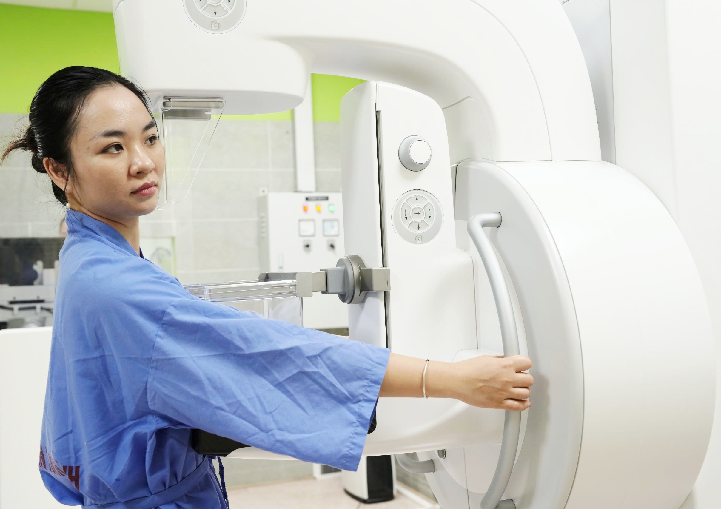
Experts state that mammography remains the most effective screening method for the early detection of breast cancer, even when lesions are only a few millimeters in size or cannot yet be detected through physical examination.
Breast Cancer Does Not Appear “Overnight”
According to physicians, breast cancer results from a silent transformation process that may last for many years within breast cells.

Initially, the cells simply undergo abnormal proliferation, known as ductal hyperplasia. Over time, as genetic mutations continue to accumulate, these lesions may progress into carcinoma in situ and eventually invasive cancer capable of spreading to other organs. This process is influenced by hormones, genetic factors, and environmental conditions.
Because the disease develops silently, detecting it at an early stage is particularly important. Early diagnosis offers patients a better chance of effective treatment, reduces the likelihood of total mastectomy, and significantly improves quality of life and long-term prognosis.
Why Is Mammography Considered the “Gold Standard” in Screening?
Mammography is a low-dose X-ray technique used to examine breast tissue structure. This method can detect extremely small lesions, especially microcalcifications — an early sign that may appear even before a distinct tumor forms.
In clinical practice, patients are usually imaged in standard positions for both breasts so physicians can comprehensively evaluate breast tissue and compare both sides.
One major advantage of mammography is that it is quick, minimally invasive, cost-effective, and suitable for widespread community screening. The images are stored, enabling physicians to monitor changes in breast tissue over time and detect subtle abnormalities early.
Many women worry that X-rays may be harmful due to radiation exposure. However, experts confirm that the radiation dose used in mammography is very low and remains within safe limits. The benefits of early cancer detection far outweigh the risks associated with radiation exposure.
Not Everyone Has the Same Risk
Current recommendations emphasize that breast cancer screening should be based on risk stratification rather than applying the same approach to all women.
For women at average risk - meaning they have no personal history of breast cancer, no high-risk genetic mutations, and no close relatives diagnosed at an early age — many current guidelines recommend beginning routine mammography screening at age 40.
Meanwhile, women at high risk, such as carriers of BRCA1 or BRCA2 mutations, those with multiple relatives diagnosed with breast cancer, or those who underwent chest radiation at a young age, require earlier surveillance, sometimes beginning between ages 25 and 30. In such cases, mammography is often combined with magnetic resonance imaging (MRI) to improve lesion detection, particularly in younger women with dense breast tissue.
Physicians warn that many women only seek medical attention after discovering a lump or experiencing persistent breast pain. However, by the time symptoms become apparent, the disease may already have progressed to a more advanced stage.
In addition to regular screening, women should pay attention to warning signs such as:
A hard lump in the breast or armpit
Abnormal nipple discharge
Thickened, red, dimpled, or distorted breast skin
Retracted nipples or changes in nipple shape
When these symptoms occur, patients should promptly visit a specialized medical facility for examination and mammography.
Detecting Cancer from the Smallest Signs
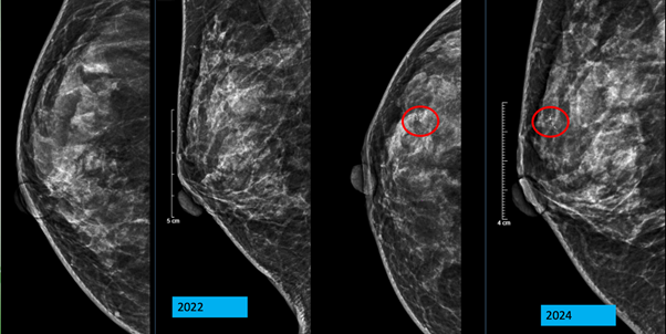
A typical case recorded at Bệnh viện Bạch Mai involved a 53-year-old woman who underwent routine breast cancer screening.
While her mammogram in 2022 showed no abnormalities, the 2024 examination revealed a small cluster of microcalcifications in the right breast. A subsequent biopsy confirmed ductal carcinoma in situ (DCIS), a very early stage of breast cancer.
According to experts, this represents the greatest value of screening: detecting disease before symptoms appear and before tumors grow large or metastasize.
Currently, at Bạch Mai Hospital, the digital mammography system provides high-quality imaging with low radiation doses. Images are stored on the PACS system and independently interpreted by two breast imaging specialists using a “double reading” process, thereby increasing the detection rate of subtle lesions and reducing the risk of missed diagnoses.


78 Giai Phong Street, Kim Lien Ward, Hanoi City
1900.888.866
096.985.1616
Department of On-Demand Medical Examination:
Outpatient Department: Monday – Friday
Main responsible person: Associate Professor, Dr. Dao Xuan Co - Hospital Director